


About Us
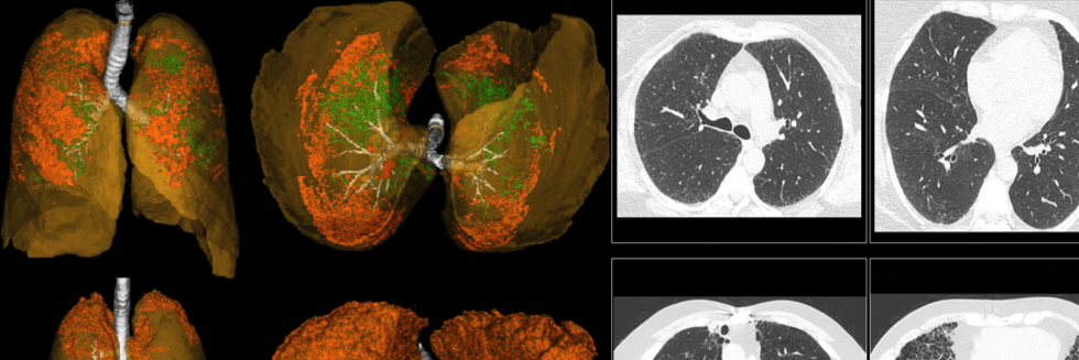
The Applied Chest Imaging Laboratory at Brigham and Women's Hospital, Harvard Medical School, is a multidisciplinary team dedicated to advancing quantitative research in the field of lung diseases. Our primary focus lies in the investigation of Chronic Obstructive Pulmonary Disease, Pulmonary Vascular Disease, and Interstitial Lung Disease. We are committed to harnessing the potential of cutting-edge imaging technology to deepen our understanding and quantification of disease processes through hypothesis-driven modeling. Our team of experts collaborates to create state-of-the-art imaging analysis and modeling algorithms, ultimately empowering clinical and genetic research to revolutionize patient care.
Our Projects
Recent Publications
- Association of outdoor temperature with lung function in a temperate climate
- Arterial Vascular Pruning, Right Ventricular Size, and Clinical Outcomes in Chronic Obstructive Pulmonary Disease. A Longitudinal Observational Study
- A graph-cut approach for pulmonary artery-vein segmentation in noncontrast CT images
- Imaging Advances in Chronic Obstructive Pulmonary Disease. Insights from the Genetic Epidemiology of Chronic Obstructive Pulmonary Disease (COPDGene) Study
- Quantitative CT Evaluation of Emphysema Progression over 10 Years in the COPDGene Study
- Equipoise in Appropriate Initial Volume Resuscitation for Patients in Septic Shock With Heart Failure: Results of a Multicenter Clinician Survey